Histology
Started June 19, 2019
138
0
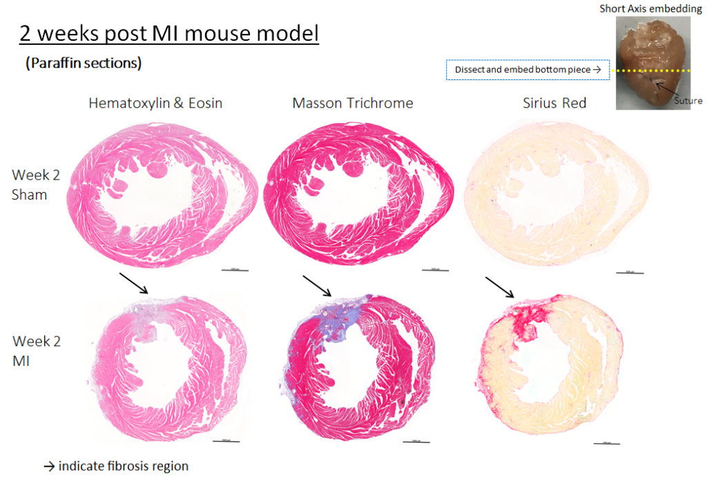
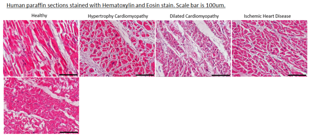
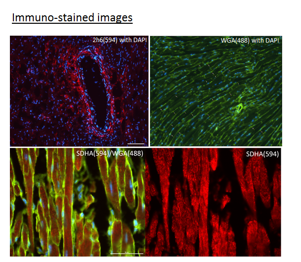
Our studies with mouse and human hearts frequently require detailed histological examination. Hearts are routinely processed into either cryo or paraffin blocks. Thin tissue slices of 4-5μM thickness are sectioned using either a cryostat or a microtome and affixed onto glass slides. Histological and immunological stainings are carried out to obtain data to assess the severity of myocardial fibrosis, cardiomyocyte cell widths for hypertrophy, or inflammatory cellular infiltrates. Whenever a new antibody or staining protocol is introduced, it is important to carry out optimisation trials prior to staining our actual samples.


Add Your Comment